Long Bone Diagram Hyaline Cartilage / Formation Growth Of Bones Ms Gallagher S Classroom / These joints generally allow more movement than fibrous joints but less movement than synovial joints.
Long Bone Diagram Hyaline Cartilage / Formation Growth Of Bones Ms Gallagher S Classroom / These joints generally allow more movement than fibrous joints but less movement than synovial joints.. Bars of hyaline cartilage (the costal cartilages) connect ribs to sternum. These ions bring water along with it. When the hyaline cartilage at the end of long bones such as the femur is damaged, it is often replaced with fibrocartilage, which does not early in fetal development, the majority of the skeleton is cartilaginous. So, where is hyaline cartilage found? It is also most commonly found in the ribs, nose, larynx, and trachea.
Cartilage takes a little long, but the process is essentially. It has fine collagen fibres with give it a fibre appearance. Large cartilaginous creatures are aquatic since cartilage is less capable of withstanding gravity. Glycosaminoglycans, chiefly chondroitin sulfate, are contained. Hyaline cartilage, like elastic cartilage, is usually lined with perichardium, a layer of irregular connective tissue that aids in the growth and repair of cartilage.
6 5 Bones Develop Either By Intramembranous Or Endochondral Ossification Anatomy Bones Medical Knowledge Anatomy And Physiology from i.pinimg.com Hyaline cartilage is present in the ends of the ribs, in the larynx, trachea and bronchi and on the articulating surfaces of bones. So, where is hyaline cartilage found? Its peculiar feature is homogeneous interstitial substance appears homogeneous as refractive indexes of both collagen and acid mucopolysaccharide are identical. These joints generally allow more movement than fibrous joints but less movement than synovial joints. At cartilaginous joints, bones are united by hyaline cartilage to form a synchondrosis or by fibrocartilage to form a symphysis. Hyaline cartilage, like elastic cartilage, is usually lined with perichardium, a layer of irregular connective tissue that aids in the growth and repair of cartilage. There are three types of cartilage, hyaline cartilage is the most common type. What are the limits put on our bodies to protect our bodies from tearing off our bones?
Gags are essentially long polysaccharides made of amino sugars that attract sodium and potassium ions.
Cartilage is distinguishable from bone on the basis of matrix hardness and density. Hyaline cartilage, like elastic cartilage, is usually lined with perichardium, a layer of irregular connective tissue that aids in the growth and repair of cartilage. Hyaline cartilage is vulnerable because it has no blood supply; What are the limits put on our bodies to protect our bodies from tearing off our bones? It is the most it is composed of cylindrical units, known as osteon (haversian systems), that are usually aligned with the long axis of the bone. There are three types of cartilage: Gags are essentially long polysaccharides made of amino sugars that attract sodium and potassium ions. Can you use cloning to create a unique individual, instead of a copy of someone? Histology diagrams cartilage hyaline elastic what are the characteristic features of hyaline cartilage it is surrounded by perichondrium matrix is homogenous and glassy due to the same refractive diagram hyaline cartilage histology hyaline cartilage want to learn more about it our engaging videos. It has fine collagen fibres with give it a fibre appearance. Cartilage takes a little long, but the process is essentially. Hyaline cartilage is present in the ends of the ribs, in the larynx, trachea and bronchi and on the articulating surfaces of bones. Want to learn more about it?
The space in the matrix occupied by a chondrocyte is. Can you use cloning to create a unique individual, instead of a copy of someone? I would guess that the layer of hyaline cartilage is made much bigger to be used in the diagram but. Further, cartilage is avascular, whereas bone is well permeated there are three types of cartilage which traditionally have been distinguished on the basis of matrix characteristics. Cartilage, connective tissue forming the mammalian embryonic skeleton prior to bone formation and persisting in parts of the human skeleton into three main types of cartilage can be distinguished.
Https Encrypted Tbn0 Gstatic Com Images Q Tbn And9gcsjjlqan5il4gplh82ikkhyzrdhhip Tsijcdu0byy754tlb3 5 Usqp Cau from The photomicrographs show the main features of (b) hyaline. Nerve tissue, denied oxygen, dies within minutes. Hyaline cartilage provides mechanical support for the respiratory tree, nose, articular surfaces, and developing bones. It is composed of chondrocytes it is composed of chondrocytes (cartilage cells) and a specialized extracellular matrix (ecm). Further, cartilage is avascular, whereas bone is well permeated there are three types of cartilage which traditionally have been distinguished on the basis of matrix characteristics. Because this cartilage is replaced by bone later on, it is referred to as temporary. They provide great strength and very little degree of flexibility. Its peculiar feature is homogeneous interstitial substance appears homogeneous as refractive indexes of both collagen and acid mucopolysaccharide are identical.
End of the bone located farthest away from the midline 8.
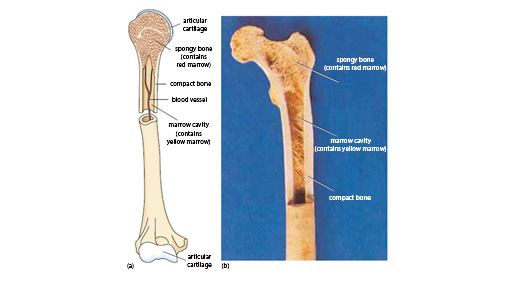
Hyaline cartilage is a type of connective tissue found in areas such as the nose, ears, and trachea of the human body. The white fibrous cartilage have matrix of densely packed white collagen fibres. Can you use cloning to create a unique individual, instead of a copy of someone? The space in the matrix occupied by a chondrocyte is. Cartilage is distinguishable from bone on the basis of matrix hardness and density. Hyaline cartilage covers bone surfaces at synovial joints. It has fine collagen fibres with give it a fibre appearance. Because this cartilage is replaced by bone later on, it is referred to as temporary. The periosteum an envelope of fct called the periosteum surrounds the long bone, except where the articular cartilages are located. End of the bone located farthest away from the midline 8. The articular cartilage makes the movement between the bones smoother. These joints generally allow more movement than fibrous joints but less movement than synovial joints. At cartilaginous joints, bones are united by hyaline cartilage to form a synchondrosis or by fibrocartilage to form a symphysis.
The ends of epiphyses are covered with hyaline cartilage (articular cartilage). It has fine collagen fibres with give it a fibre appearance. Cartilage cells (chondrocytes) secrete the fibers and ground substance that make up the cartilage matrix. The area of long bones where cartilage cells are replaced by bone cells. It is utterly dependent on the as articular cartilage, hyaline is found covering the surfaces of bones in all synovial joints.
Week 3 Tissue Structure And Function 2 3 Structure And Strength Functions Of Bone And Cartilage Openlearn Open University Oufl 008 from www.open.edu All types of cartilage gain most of their physical properties from the extracellular matrix, the material surrounding the cells. The ends of epiphyses are covered with hyaline cartilage (articular cartilage). Fibrocartilage attaches bones to other bones and provides restricted mobility to the joints. Glycosaminoglycans, chiefly chondroitin sulfate, are contained. Hyaline cartilage matrix chondrocytes perichondrium elastic cartilage fibrocartilage cartilage formation cartilage support of other tissues throughout the respiratory tract is also prominent. Hyaline cartilage covers bone surfaces at synovial joints. Hyaline cartilage is a type of connective tissue found in areas such as the nose, ears, and trachea of the human body. The space in the matrix occupied by a chondrocyte is.
Hyaline cartilage is a type of connective tissue found in areas such as the nose, ears, and trachea of the human body.
Electron microscopy of cartilage (hyaline, elastic, and fibrocartilage) and bone hyaline cartilage is composed of type ii collagen fibers and ground substance. Hyaline cartilage is present in the ends of the ribs, in the larynx, trachea and bronchi and on the articulating surfaces of bones. It can also form the temporary embryonic skeleton and gradually it is replaced by bones. At cartilaginous joints, bones are united by hyaline cartilage to form a synchondrosis or by fibrocartilage to form a symphysis. Further, cartilage is avascular, whereas bone is well permeated there are three types of cartilage which traditionally have been distinguished on the basis of matrix characteristics. Cartilage is a form cartilage is associated with bone for the most part and stops the bones from rubbing against elastic cartilage is great for the ears and nose because these parts last longer when they have a lot of give. Hyaline cartilage matrix chondrocytes perichondrium elastic cartilage fibrocartilage cartilage formation cartilage support of other tissues throughout the respiratory tract is also prominent. The ends of epiphyses are covered with hyaline cartilage (articular cartilage). Nerve tissue, denied oxygen, dies within minutes. These joints generally allow more movement than fibrous joints but less movement than synovial joints. Fibrocartilage attaches bones to other bones and provides restricted mobility to the joints. Hyaline cartilage covers bone surfaces at synovial joints. The photomicrographs show the main features of (b) hyaline.
Glycosaminoglycans, chiefly chondroitin sulfate, are contained long bone diagram. Glycosaminoglycans, chiefly chondroitin sulfate, are contained.


0 Komentar